What Does Babyecho Mean?

For most ladies, ultrasound reveals that the child is expanding typically. If your ultrasound is typical, just be sure to keep going to your prenatal check-ups. Sometimes, ultrasound may show that you and your baby requirement unique treatment. As an example, if the ultrasound shows your baby has spina bifida, he may be dealt with in the womb before birth.
A c-section is surgical procedure in which your baby is birthed with a cut that your doctor makes in your stomach and uterus. Whatever an ultrasound shows, talk with your copyright concerning the most effective look after you and your child - baby doppler. Last examined: October, 2019
During this check, they will check the baby is expanding in the ideal area, whether there is greater than one infant and they will certainly likewise examine your baby's growth thus far. This screening is readily available in between 10 14 weeks of maternity and is made use of to assess the possibilities of your child being born with several of these problems.
The Babyecho Statements
It entails a combined test of an ultrasound scan and a blood test. During the scan, the sonographer will measure the fluid at the back of the baby's neck to figure out 'nuchal clarity' - https://papaly.com/categories/share?id=3134c5f975bd4b22ba97f387cd893054. They will certainly after that determine the opportunity of your child having Down's, Edwards' or Patau's disorder using your age, the blood test and check outcomes
During this scan, the sonographer checks for architectural and developing irregularities in the infant. During this scan consultation, you might be used screenings for HIV, syphilis and hepatitis B by an expert midwife. Sometimes, a third-trimester scan is recommended by your midwife adhering to the outcomes of previous examinations, previous issues or existing clinical conditions.
The standard 2D ultrasound creates level and detailed images which can be used to see your baby's internal body organs and aid find any type of interior problems. These black and white photos assist the sonographer identify the baby's pregnancy, development, heartbeat, advancement and size. Some pregnant moms pick to have a 3D ultrasound scan due to the fact that they reveal more of a real-life picture of the baby.
Babyecho - An Overview
3D ultrasound scans show still photos of your child's outside body as opposed to their insides, so you can see the shape of the infant's face features. 4D ultrasound scans are similar to 3D scans yet they show a moving video clip as opposed to still images. This catches highlights and darkness better, consequently creating a more clear picture of the baby's face and activities.
:max_bytes(150000):strip_icc()/191127-ultrasound-trimester-pink-2000-fd089add04f8444e9d7a403933d1994f.jpg)
A is detected throughout this check. Most moms and dads choose for this scan for.
The Of Babyecho
Sometimes a may be required to obtain and a more clear picture. This is generally performed and occasionally a may be required (heart doppler). Validate that the child's heart is present; To much more accurately.
Please see below. It coincides as 19-22 weeks, but some may be or in the and it may to. Generally this is used if there are such as visit our website spina bifida or if moms and dads are eager to understand the earlier. These scans may be done, however a few of the and thus, a is required to This scan is done generally at.
4 Simple Techniques For Babyecho
:max_bytes(150000):strip_icc()/191127-ultrasound-trimester-pink-2000-fd089add04f8444e9d7a403933d1994f.jpg)
Furthermore, the can be by by an. and is monitored by these scans. of, andare done to get to an. around the baby is determined. and child's are examined. () The way nearer the is beneficial to. Occasionally, an which was previously might be.
10 Simple Techniques For Babyecho
If, these scans might be to. (of the infant) can likewise be executed. This consists of, along with; This consists of, along with (14-20 weeks).
A scan is vital before this test is done.
9 Simple Techniques For Babyecho
The examination can offer important information, aiding women and their health-care providers handle and care for the pregnancy and the unborn child.
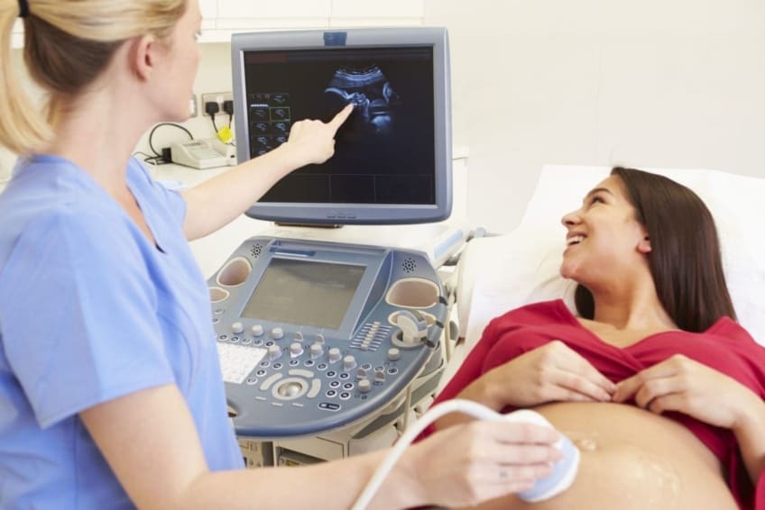
A transducer is placed right into the vaginal area and rests versus the back of the vaginal canal to create a picture. A transvaginal ultrasound produces a sharper photo and is commonly used in early pregnancy. Ultrasound makers are about the dimension of a grocery store cart. A TV screen for checking out the images is affixed to the machine (https://pastebin.com/u/babydoppler1).
Comments on “How Babyecho can Save You Time, Stress, and Money.”